
How molecular imaging helps locate prostate tumours
PET-CT PSMA helps accurately detect lesions in prostate cancer, guiding treatment and identifying potential recurrences.
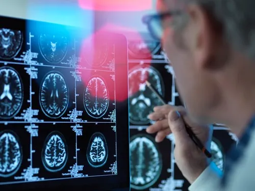
 Foto ilustrativă
In prostate cancer, pinpointing the exact location of the disease can play an important role in choosing the most appropriate treatment. Modern imaging technologies now allow tumour cells to be identified based on their specific biological characteristics.
One such method is PSMA PET-CT, which uses a radioactive substance (radiotracer) that binds to PSMA — a protein found in large quantities on the surface of many prostate cancer cells. The PET component of the scan shows the areas where this tracer accumulates, whilst the CT component provides precise anatomical information about the location of these areas. By combining the two techniques, doctors can detect lesions both in the prostate or in the region where it was surgically removed, as well as in the lymph nodes, bones, or other organs, thereby gaining a clear picture of how far the disease has spread.
This investigation is used to stage the disease in certain patients, as well as to identify a possible recurrence when PSA levels begin to rise following completion of the initial treatment. The information obtained can influence clinical decisions regarding radiotherapy, surgery, or systemic treatments. In advanced cases of the disease, identifying PSMA-expressing lesions can also help select patients who are eligible for targeted therapy with lutetium-177.
Content paraphrased and adapted by SeniorHelp from verified public sources.
Original source: Realitatea →Previous article
486 communes in Romania have no pharmacy. How the elderly get their hands on medication
Next article
Healthy digestion: which snacks aid bowel transit and which foods cause bloating
Similar news

The biological age of the heart, estimated with AI, can indicate the risk of death in people over 50
14 November 2026

Harvard Study: for a longer life, variety of exercise matters, not just movement
14 November 2026

Diabetes: over 40% of sufferers don't know they are affected. The signs that must not be ignored
14 November 2026
Type 2 diabetes increases the risk of Alzheimer's by 53%, a new study shows
14 November 2026
 Foto ilustrativă
How Much Salt Is It Healthy to Eat Daily. What a Cardiologist Says About the Risks and Foods with Hidden Sodium
14 November 2026

Ministry of Health launches the 2027–2031 Digitalisation Strategy: a single electronic record for patients
13 November 2026